
This web page represents a distillation of the major ideas and information associated with the Biological Science (BIOL 1003) lecture course. References to page numbers, Figures, and Tables, are associate with reading assignments Asking about Life (3rd edition; 2005) by Tobin and Dusheck - reading assignments are noted in the Syllabus for BIOL 1003.003 for additional information. If you note any errors in the following document, I'd appreciate it if you would bring this to my attention. Email address: mhuss@astate.edu.
LECTURE EXAM II IS SCHEDULED FOR WEDNESDAY, MARCH 15TH. IF YOU WOULD LIKE TO TRY YOUR HAND AT A PRACTICE TEST FOR THE SECOND SET OF MATERIAL THAT WE HAVE COVERED THUS FAR IN THE SEMESTER, ACCESS THIS INFORMATION BY VISITING THE FOLLOWING LINKS. KEEP IN MIND THAT THIS NOT THE ACTUAL EXAM BUT IT OUGHT TO GIVE YOU A FEEL FOR THE KINDS OF QUESTIONS THAT MIGHT BE ASKED ON THE ACTUAL "IN CLASS" EXAM ON MARCH 15TH.
- PRACTICE EXAM II
- PRACTICE EXAM II - ANSWER KEY (THE CORRECT RESPONSE IS HIGHLIGHTED IN YELLOW AND THE FONT IS BOLDED)
- Also check out the ANSWER KEY TO MITOSIS WORKSHEET.
Cell Crossword Puzzle: PDF file (Optional "Bonus" Assignment: Due February 24, 2006)
Cell & Cell Division Vocabulary Words and Terminology: MS WORD FILE (Optional "Bonus" Assignment: Due March 1, 2006)
A HIERARCHY EXISTS COMPOSED OF DIFFERENT LEVELS OF ORGANIZATION WHICH ALLOWS LIFE AS IT CURRENTLY EXISTS TO PERSIST ON EARTH:
ATOMS ===> MOLECULES ===> ORGANIC MACROMOLECULES===>CELLULAR COMPONENTS ===> CELLS (fundamental unit of life for single-celled microbes to multicellular life forms) ===> TISSUES ===> ORGANS ===> ORGAN SYSTEM ===> MULTICELLULAR ORGANISM (e.g., plants, animals, and fungi)===> POPULATION (SAME SPECIES) ===> COMMUNITY (DIFFERENT SPECIES) ===> ECOSYSTEM (COMMUNITIES AND THEIR ENVIRONMENT) ===> BIOSPHERE (ALL LIFE ON EARTH)
The fundamental unit that possesses all of these characteristics of life is found in the cell. We recognize the cell as being alive!
- Robert Hooke, 17th century (1665) - dead cells of cork and coined the word "cell" after a name which referred to a small room (monastery, convent, or prison cell).
- Anton van Leeuwenhoek - 17th century (1675), developed first light microscopes and saw the first living cells, which he called animalcules - "little animals". Refer to Figure 4-1 on page 65 in the textbook.
- 1839 - Matthias Schleiden and Theodor Schwann proposed that all living animals and plants consist of cells. Their work and others lead to the CELL THEORY (i.e., all organisms, except viruses, are made up of cells).
- Virchow in the mid 19th century - cells give rise to new cells.
- What kind of
cells are there?
- Prokaryotic cells: Nucleus absent - cells are 0.4-5.0 micrometers or microns
(1 micron = one millionth of a meter in distance) in size.
- Single-celled organisms - bacteria, "blue-green" algae or cyanobacteria.
- Eukaryotic
cells: Nucleus present - cells are 10-100 micrometers in size.
- Single-celled organisms - protists, amoebae, paramecia, unicellular green algae.
- Colonial organisms - volvox, slime molds.
- Filamentous organisms - Spirogyra (pond scum, green alga), fungi. Multicellular organisms - differentiated cells take on special functions: animals, plants, and fungi.
- Prokaryotic cells: Nucleus absent - cells are 0.4-5.0 micrometers or microns
(1 micron = one millionth of a meter in distance) in size.
I. Discovery of cells, the microscope and cell theory
In 1665, Robert Hooke using a crude light microscope saw and described cells in a piece of cork. This is a good example of how certain discoveries are dependent on the development of appropriate technology. The discovery of cells was dependent on the development of the light microscope. Can you think of any other examples where this is true? [Laboratory equipment and procedures (e.g., refrigeration, electrophoresis, ultracentrifugation, etc...) required for many types of scientific study would not be possible without a grasp of basic technology, such as, the production and use of electricity, which has only been around for about one century; Detailed discoveries about our solar system were dependent on development of rocket propulsion systems, computers, educated and technologically sophisticated support personnel, etc.]
Three ways to make visual observations - Refer to Extreme Biology on page 66 in textbook on details of microscopy:
A. Unaided eye
B. Light microscope (living and nonliving material)
C. Electron microscopea. transmission electron microscope (specimens are sliced thin, treated with chemicals that allow some parts of specimen to deflect electrons, while others allow the beams to pass through the object).
b. scanning electron microscope (object covered with carbon, platinum, or gold), causing the electron beam to bounce off the object, giving a 3D appearance to object).
- magnification - the process of increasing the apparent size of an object.
- resolution - the ability to separate two or more objects that are close together and show them as distinct entities.
- contrast - an objects ability to absorb more or less light than its surroundings (contrast increased by use of stains and phased light). Which is more important? Resolution, it is useless if you magnify something a thousand times if it appears fuzzy.
- Resolving power of a light microscope is about 0.2 micrometer or microns (Ám) or 200 nanometers (nm). If distance between two objects is less than 0.2 Ám, we see them as a single object. Resolving power of electron microscope is about 2 nm or about 100 times greater resolution than is possible with the best light microscope.
II. Components of the prokaryotic and eukaryotic cell
Cells are the smallest unit of life, because they have complex organization, metabolic activity, and reproductive behavior.
Prokaryotic cells or prokaryotes - bacteria and cyanobacteria (AKA "blue-green algae), possess no true membrane-bound nucleus or other membrane-bound organelles. The genome (genetic makeup) of prokaryotes is represented by a circular loop of naked DNA (nucleoid region). Ribosomes (comprised of two subunits of a protein/rRNA complex) are found in the cytoplasm of the cells. The cell is bounded by a membrane and a cell wall. Depending on species, some prokaryotes possess one flagellum or multiple flagella (making it possible for them to swim). Some bacteria are surrounded by a slimy polysaccharide capsule.
Refer to diagram of a "typical" bacterial cell: In textbook in Figure 4-2B on page 68 and Figure 20-6 on page 417 (DON'T READ CHAPTER 20, JUST REFER TO THIS PARTICULAR DIAGRAM) AND VISIT http://www.cellsalive.com/cells/bactcell.htm.
- Eukaryotic cells or eukaryotes - single-celled protists (refer to Figure 4-3 on page 69 in the textbook), such as amoebae and green algae, and multicelluar organisms, such as plants, animals, and fungi.
- Eukaryotes have cells which possess a "true nucleus" and other membrane-bound organelles. Each membranous sac or organelle performs a specific duty in the body of the cell. Some of the organelles are thought to have occurred as a result of a symbiosis (mutually beneficial relationship) between a prokaryote and a eukaryote (Lynn Margulis - Endosymbiont Theory on the origin of chloroplasts and mitochondria).
- Refer to diagram of a "typical" animal cell presented in class and in Figure 4-5 on page 73 in the textbook AND visit http://www.cellsalive.com/cells/animcell.htm.
- Parts of a
typical eukaryotic cell or eukaryote and their function
- Plasma or cell membrane is semi-permeable (channel and carrier proteins on the surface or embedded in the membrane are responsible for active and passive transport). Phospholipid bilayer membrane - follows the fluid mosaic model of cell membrane structure.
- Cytoplasm or protoplasm - fluid matrix that organelles are embedded in, largely made of water and dissolved substances (proteins, sugars, salts, etc...). Also contains the cytoskeleton (network of protein fibers) that help to anchor organelles and help give the cell its shape.
- Nucleus - chromatin or chromosomes (DNA and protein) + nucleolus (nucleolus organizer region - NOR on chromosomes) which produces rRNA which is found complexed with protein in the ribosomes (protein synthesis). Bound by membrane - nuclear envelope which contains pores.
- Endoplasmic reticulum - rough (with ribosomes) and smooth (without ribosomes) - channels of membranes involved as site of protein and lipid synthesis and movement of protein products.
- Golgi apparatus - storage of proteins, lipids, and carbohydrates.
- Mitochondrion - power house of the cell - aerobic respiration. Flagella and Cilia are made up of microtubules in a 9 + 2 arrangement - movement.
- Lysosomes and peroxiosome - membranebound vesicles which contain enzymes for the breakdown of particles and toxic compounds (the latter detoxifies hydrogen peroxide).
Plant cells
are the same as animal cells, except
these possess a cell wall made up of cellulose fibrils and pectin (glue-like
polysaccharide), plastids (e.g., chloroplasts - associated with photosynthesis,
leucoplasts - starch or oil storage, chromoplasts - pigment-containing), and
large vacuoles. Plants lack centrioles, which occur in pairs, composed of
microtubules. Refer to diagram of a
"typical" plant cell, presented in class, in the diagram below, and in Figure
4-5 on page 72 in the textbook AND visit
http://www.cellsalive.com/cells/plntcell.htm.
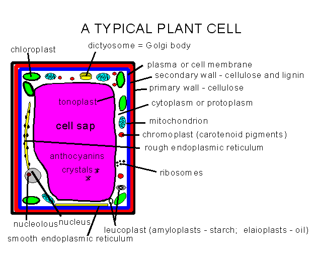
OR VISIT THE FOLLOWING LINK AT http://sun.menloschool.org/~cweaver/cells/
Cells of
fungi
are more similar in their basic structure to animal cells than these are to
plant cells. Fungal cells do possess cell walls which may contain
cellulose, but these are more likely to possess the polysaccharide called
chitin. Fungal and animal cells both possess centrioles, sterols in their
cell membranes, glycogen (as a food storage molecule; plants use sucrose and
starch), bioluminescence found in both groups, and both are heterotrophs or
"other feeders" (unable to make their own food).
- To review slide presentation on the material presented above, click onto either of the following hyperlinks for more information:
SEE TABLE 4-1 on page 71 for a summary of cell features common and distinctive among different major groups of organisms.
Also take a look at:
CELLS AS ART: http://thalassa.gso.uri.edu/flora/arranged.htm
MEMBRANE STRUCTURE AND FUNCTION
Membrane Structure
Biological membranes are made up of a phospholipid (hydrophobic tails - fatty acids and hydrophilic heads - phosphate group) bilayer and proteins (peripheral and integral or transmembrane) proteins ==> Fluid mosaic model of membrane structure (refer to page 90, Figures 4-18 in the textbook)
If fatty acid tails are unsaturated they become bent and this increasing the fluidity of the membrane making it more unstable (refer to Figure 4-15 on page 82, Figure 3-9C on page50 in the textbook). Other lipids, such as cholesterol (animals) and ergosterol (fungi) become integrated into the membrane.
Properties of biological membranes
- membranes are oily and can change structure by sliding the phospholipid layers over one another.
- membranes are different on one side than on the other - this contributes to ASYMMETRY.
- proteins attached to carbohydrates = glycoproteins are involved in cell to cell communication and enhancing asymmetry because of their position on the outer side (exoplasmic face) of the cell membrane.
- membranes are semipermeable or differently permeable (large uncharged polar molecules like glucose, and ions don't pass through membrane, but hydrocarbons, water, oxygen and carbon dioxide go straight through).
- Some function of membranes
- movement of water and solutes into and out of cells and organelles.
- differential permeability.
- cellular communication - binding of certain chemicals to cell membrane, initiate metabolic changes inside cell.
- Movement of water and other molecules across membranes
- diffusion - random movement of molecules in a fluid or gas from a region of high concentration to a region of low concentration. [EXAMPLES: dye in water, perfume in the air]
- diffusion occurs along a concentration gradient. To go against a concentration gradient always requires the input of energy.
- Refer to Figure 4-19 on page 86 in the textbook.
OSMOSIS
Thought experiment: A beaker containing 2% sucrose is gently placed into a larger beaker containing distilled water. Sucrose will tend to diffuse out of the small beaker into the larger beaker along a concentration gradient. Water will also tend to diffuse into the beaker from a region of high concentration (surrounding distilled water) to a region of low concentration (the water displaced by the presence of a solute - sucrose).
If you place a semipermeable membrane (i.e., permeable to water, but impermeable to sucrose) over the surface of the small beaker, then sucrose cannot cross the barrier but water can! Osmosis is the movement of water across a differentially permeable membrane along a concentration gradient. Refer to Figure 4-16 on page 83 in the textbook.
TONICITY (tonos = tension)
- Hypotonic - low solute concentration relative to another solution.
- Hypertonic - high solute concentration relative to another solution.
- Isotonic - solute concentration is the same as that of another solution.
The use of these terms is always in relationship to another solution. For example:
Distilled water =========>2% NaCl ==============> 5% NaCl
hypotonic =============> hypertonic
hypotonic ============> hypertonic
hypotonic
===================================> hypertonic
TURGOR PRESSURE - Water moving into cell pushes the cell membrane up against the cell wall causing turgor pressure. Loss of water from the vacuole/cytoplasm causes shrinkage of cellular contents, or PLASMOLYSIS (Refer to Figure 4-18 on page 85). Loss of water from plant cells results in wilted tissue!
Red blood cells or RBCs or erythrocytes (Refer to Figure 4-17 on page 85).
- If external solution is isotonic (0.9 % sodium chloride solution) ==> no change in cell volume.
- If external solution is hypertonic ==> cells shrivel (Hence the saying, if lost at sea with no drinkable water, "Water water everywhere, but not a drop to drink!").
- If external solution is hypotonic ===> cells swell and explode!
TRANSPORT
PROTEINS
Facilitated
diffusion
(refer to Figure 4-19A and 4-19B on page
86) occurs by means of a channel or carrier
protein, gate open and closes, or gate stays continuously open. Process works
along a concentration gradient and requires no input of energy ==> PASSIVE
TRANSPORT!.
ACTIVE TRANSPORT ==> transport of a substance across the membrane against the concentration gradient and requires the input of energy (high energy bond from ATP). Refer to Figure 4-22C on page 86.
The setup for cotransport usually requires an investment of energy by the cell. Channel protein allows one substance through that pulls another through. Both substances may move across the membrane together (symport) or in opposite directions of one another (antiport). Ion pumps use and electrochemical gradient to drive substances across the membrane.
BULK TRANSPORT OF MATERIALS ACROSS A MEMBRANE
ENDOCYTOSIS - bypasses membrane transport and allows for movement of large molecules and quantities across membrane into the cell. Refer to Figure 4-21A on page 88. Phagocytosis is form of endocytosis - occurs in amoeba and white blood cells to capture bacteria and other microbes.
EXOCYTOSIS - bypasses membrane transport and allows for movement of large molecules and quantities across membrane out of the cell. Refer to Figure 4-21B on page 88.
- Life gives rise to life (19th century notion of "Spontaneous Generation" dispelled by work of Louis Pasteur - Refer to Figure 18-3 on page 378 (DON'T READ CHAPTER 18, JUST LOOK AT FIGURE 18-3)
- The Slow Death of Spontaneous Generation (1668-1859) by Russell Levine and Chris Evers (Read about the debate in the 17th, 18th, and 19th centuries on the Spontaneous Generation of Mice, Maggots, and Microbes). Learn more about the work of Francesco Redi, John Needham, Lazzaro Spallanzani, and Louis Pasteur at http://www.accessexcellence.org/RC/AB/BC/Spontaneous_Generation.html
- A closer look at Louis Pasteur (1822-1895) by Seung Yon Rhee at http://www.accessexcellence.org/RC/AB/BC/Louis_Pasteur.html
- 1700-1900 - Two centuries of Biological Discovery - http://www.accessexcellence.org/RC/AB/BC/1750-1900.html
- Organisms give rise to other organisms.
- Cells give rise to cells.
- Cells divide so that growth and/or reproduction can take place.
Prokaryotes (i.e., bacteria, blue-green algae) are single-celled and reproduce through the process of binary fission. The circular loop of DNA (the genome) replicates and the cell divides into two equal halves. Refer to Figure 8-3 on page 153. Mitochondria and chloroplasts also reproduce by binary fission (these organelles are maternally inherited).
Eukaryotes have a nucleus and in the nucleus resides chromatin or uncondensed chromosomes. Chromatin is approximately 40 % DNA and 60% protein. See Figure 8-9 on page 160 in the textbook. DNA is tightly coiled and this coiling, packaging, and scaffolding of the DNA is facilitated by the protein. Chromatin exists as heterochromatin and euchromatin. Heterochromatin is tightly coiled, therefore the DNA is not exposed, whereas euchromatin is less tightly coiled and is exposed. Exposed DNA can be expressed to produce RNA and protein.
- Chromosomes can exist in an uncondensed or condensed state - Refer to Figures 8-7 and 8-8 on pages 158-159 in the textbook.
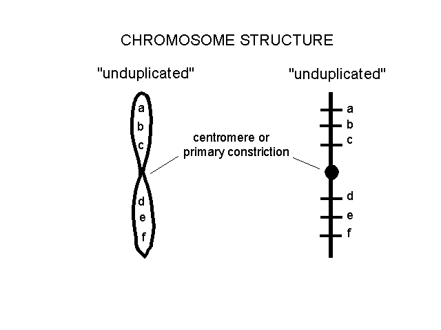
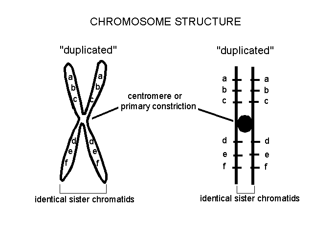
- Chromosomes can exist in an unduplicated or duplicated state (2 sister chromatids attached at the centromere.
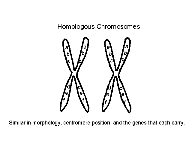
- Homologous chromosomes or homologues represent pairs of chromosomes that are similar in structure, morphology, centromere position and genes that they carry.
- Centromere = primary constriction - site of spindle fiber attachment during mitosis and meiosis.
- Chromosome number and karyotypes (arranged from biggest to smallest)
- Locus (plural - loci) - the physical location of a gene on a chromosome
- Humans possess 23 pairs of
chromosomes or a total of 46 chromosomes.
Refer to Figure
8-4 on page 154 and 9-6on pages 170.
Other examples include:
- chimpanzees (Pan) and gorillas (Gorilla) = 24 pairs of chromosomes (2N = 48)
- dogs, wolves, foxes, coyotes are all members of the genus Canis = 39 pairs of chromosomes (2N = 78)
- cats = 19 pairs of chromosomes (2N = 38)
- peas = 7 pairs of chromosomes(2N = 14)
- onions = 8 pairs of chromosomes (2N = 16)
- fruit flies = 4 pairs of chromosomes (2N = 8)
- male donkey, Equus asinus, 31 pairs of chromosomes (2N=62)
- female horse, Equus caballus, 32 pairs of chromosomes (2N=64)
- mule (the sterile offspring of a horse and donkey), 31 pairs of chromosomes + 1 lone chromosome (2N = 63).
- Most chromosomes in a set are called autosomal chromosomes (autosomes); in many organisms one pair exists that differ from autosomal chromosomes. These are called the sex chromosomes and function to control certain aspects of the gender of the individuals which possess certain combinations - Refer to Figure 9-17 on page 179 in the textbook.
Growth, replacement of dead cells, and/or asexual reproduction.
Mitosis (nuclear division) + cytokinesis (cytoplasmic division - refer to Figure 8-2 on page 151 and Figure 8-6 on page 156). No change in the genetic makeup of the individual cell (chromosome numbers remain constant).
Cell division - mitosis (Figure 8-5 on page 155 and 8-6 on page 156 in the textbook) and cytokinesis (Figures 8-9 and 8-10 on page 176 in the textbook).
G1 (gap 1 - interval before DNA replication) ====> S (synthesis - replication of DNA and synthesis of proteins found in chromatin/chromosomes) ====> G2 (gap 2 - interval before onset of mitosis)
G1, S, and G2 represent Interphase. Refer to refer to Figure 8-5, page 155 in the textbook.
Movement of chromosomes is coordinated by organized arrays of microtubules - the spindle apparatus.
Formation of haploid cells for use in producing spores or gametes.
Meiosis (reduction division) + cytokinesis (cytoplasmic division). Chromosome numbers reduced by half.
1. Diploid cell (2N) containing 2 sets of chromosomes gives rise to haploid cells (1N) containing one set of chromosomes. Meiosis is a reduction division that produces a spore OR a gamete (i.e., an egg or sperm).
2. Meiosis is divided into two parts: meiosis I (pairing of homologous chromosomes, crossing-over, and separation of homologues to separate cells); meiosis II (separation of non-identical sister chromatids into sister chromatids which go to separate poles of a dividing cell - refer to Figure 9-12 on page 175 in the textbook.). In plants and fungi, these haploid nuclei become integrated into specialized cells = spores.
Terminology and figures for understanding meiosis and related topics
- synapsis - pairing of homologues during prophase I.
- bivalent - one of the pairs of homologous chromosomes which associate during the first prophase of meiosis.
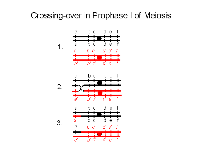
- chiasmata - the points at which homologous chromosomes remain in contact as the chromatids move apart during the first prophase of meiosis. There may be up to eight chiasmata in a bivalent pair of chromosomes. chiasma (sing.)
- gene linkage - two genes OR loci are located on the same chromosome. The closer these two genes are positioned together on the same chromosome the less likely these will appear to assort independently during meiosis. This fact allows geneticists to construct linkage maps of the genomes of different organisms, including humans. HUMAN GENOME PROJECT (FYI: A discussion of the Human Genome Project can be found in Chapter 14 in the textbook).
2 Divisions occur in Meiosis (Meiosis I and Meiosis II)
MEIOSIS I
Prophase I
- Chromatin condenses into individual chromosomes.
- Pairing of homologues begins (synapsis).
- Synapsis complete and chromosomes thicken and shorten.
- Paired chromosomes begin to repel one another => chiasmata (points along paired homologues where both are still connected). This stage is associated with crossing-over!
- Genetic recombination through crossing-over! Refer to Figure 9-11 on page 175 in the textbook.
- Maximum contraction, paired chromosomes, nuclear membrane and nuclei breakdown.
Metaphase I - chromosomes migrate to middle part of cell; spindle fibers attach to centromere; Process is random - INDEPENDENT ASSORTMENT- Refer to Figure 9-12 (upper portion of figure) on page 175 and Figure 9-13 on page 176.
Anaphase I - Homologues (i.e., parental chromosomes) separate and move to opposite poles.
Humans have 223 or 8,388,608 possible combinations of chromosomes that can end up in a gamete. With fertilization, this number is squared (8,388,608)2 = 70 trillion possible zygotes that can arise from a single mating event between two people! You're not one in a million, you're one in 70 trillion! And that's not counting the effects of crossing-over!
Illustrates the point that the purpose of meiosis is not just about reproduction or sex, its about creating novel genetic combinations.
- Crossing-over and genetic recombination
- Homologous chromosomes assort independently
- Fertilization - two different gametes fuse bring together different combination of alleles.
Telophase I - two haploid sets of chromosomes now exist, each set at the opposite pole of cell.
MEIOSIS II
Prophase II - chromosomes condense.
Metaphase II - chromosomes move to middle part of cell.
Anaphase II - separation of non-identical sister chromatids into sister chromosomes. Refer to Figure 9-12 (lower portion of figure) on page 175.
Telophase II - nuclei reform and you are left with 4 haploid nuclei.
GAMETOGENESIS IN ANIMALS LEADS TO THE FORMATION OF GAMETES
The products of meiosis must undergo differentiation in order to become functional gametes.
Spermatogenesis - 4 small sperm (cytokinesis divides the cytoplasm equally) - QUANTITY!
Oogenesis - 1 large ovum or egg and 3
small polar bodies (cytokinesis divides the cytoplasm unequally) - QUALITY!
Refer to Figures 9-7 and 9-9 on pages 171 and 173 in textbook regarding sexual reproduction.
- Polyploidy - a genetic accident that leads to more than two sets of chromosomes nucleus; lethal in most animals; more common in plants (e.g., rutabaga, wheat, many fern species are polyploids).
- Aneuploidy - consequence of nondisjunction (failure of paired homologues to separate during meiosis) leads to more or less chromosomes numbers in the resulting gametes. The fusion of a sperm and an egg results in a fertilized egg (and embryo, if viable) that contains more or less chromosomes than the "typical chromosome number" Trisomy 21 (possession of three 21 chromosomes leads to a condition called Down Syndrome - Refer to Figures 9-14, 9-15, and 14-13 on pages 177 and 313 in textbook.
|
This page was assembled by Martin J.
Huss, who can be reached at
mhuss@astate.edu.
Last revised on: MARCH 9, 2006.
