For additional
information, particularly about fungal species mentioned in the subsequent
text, also refer to the paperback, Medically Important Fungi: A Guide
to Identification by Larone.
Superficial Infections
Cellular response of host usually absent -
no pathology elicited by fungus
-
Organism remote from living tissue (primarily in the stratum corneum -
compact dead layer of skin).
-
Infection is insignificant and ignored by bodies defenses.
Identification accomplished several ways
-
Visual Inspection
-
Wood's lamp - U.V. lamp to look for presence of fluorescence.
-
Scraping off cells onto a slide and clearing tissue with 10% KOH and flaming
the slide. Look for fungal tissue (e.g., mycelial fragments, yeast
cells, spores). These slides are not permanent because the KOH causes
cells to breakdown over time - too caustic!
-
Stripping tape - Scotch tape is pulled of skin, mounted on a slide,
and stained with crystal violet, methylene blue, or iodine.
-
Isolation and subculturing of fungus.
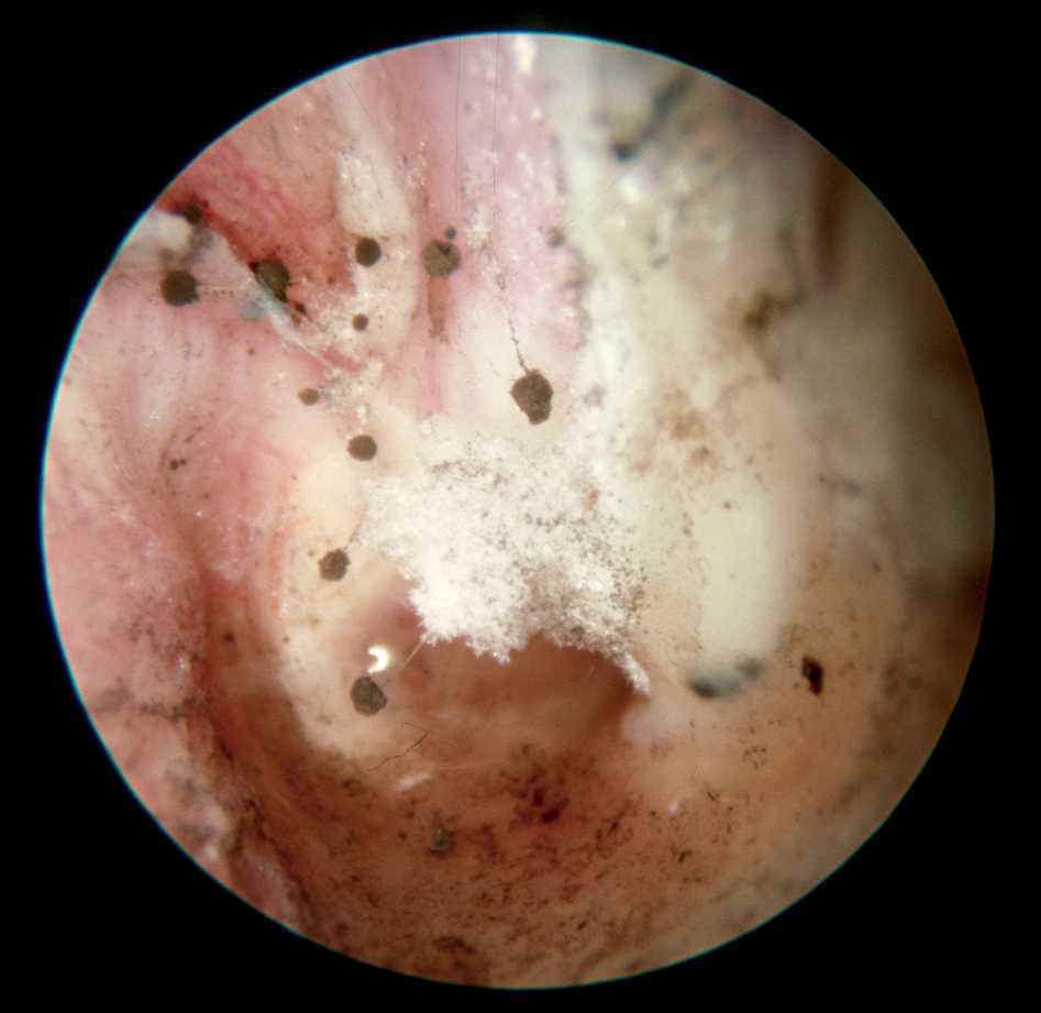
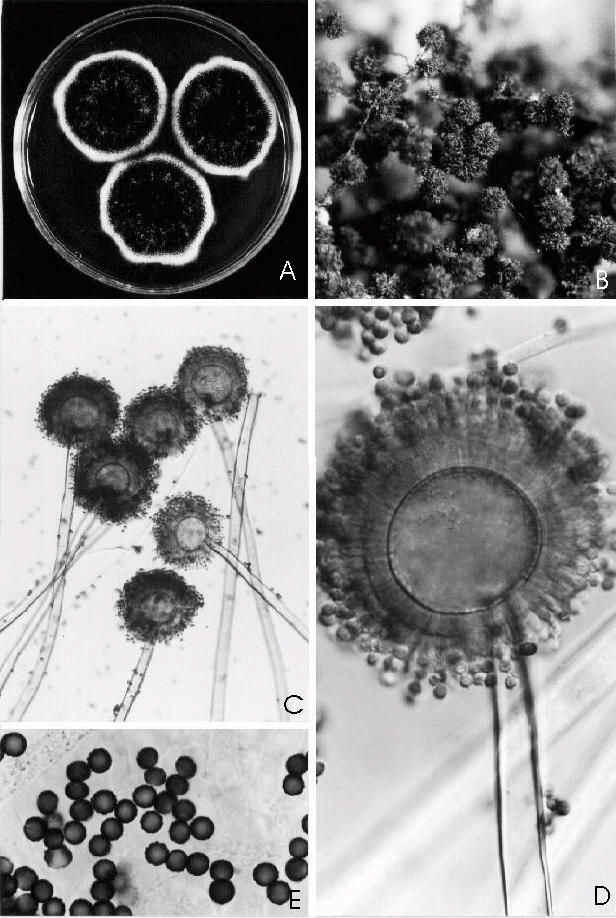
OTOMYCOSIS
Fungal Growth in the Ear Canal
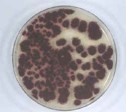
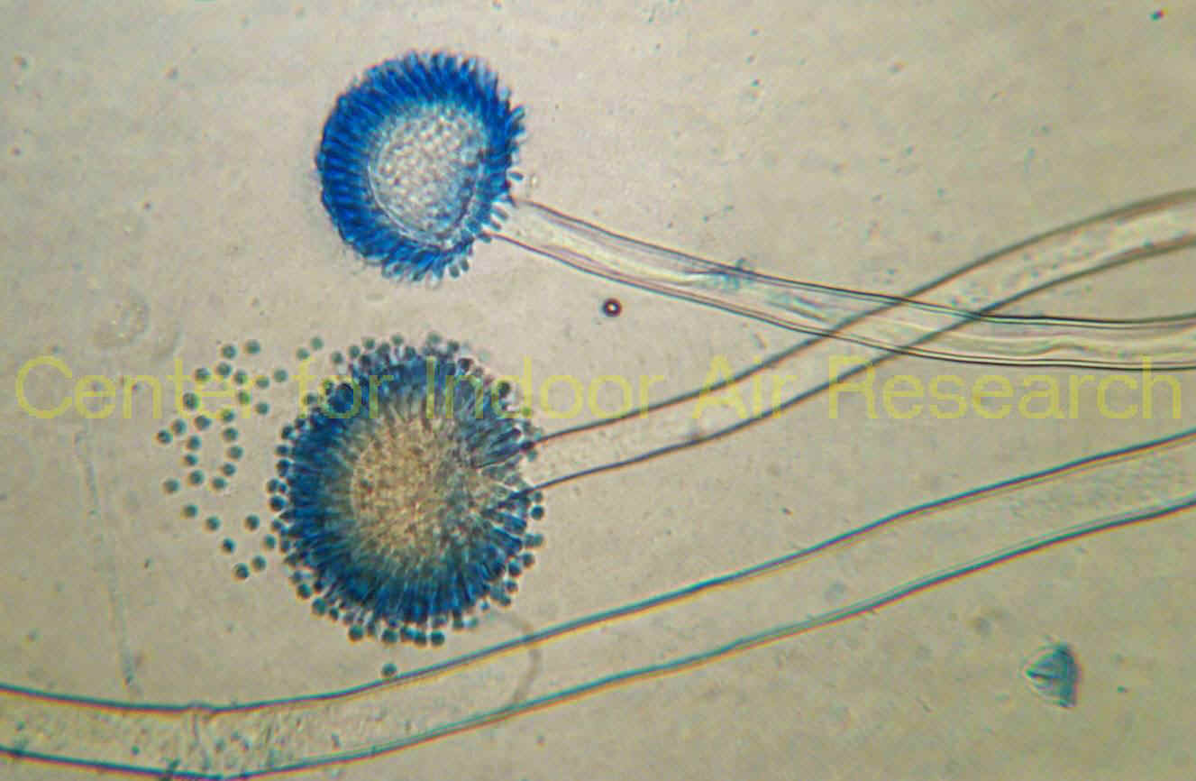
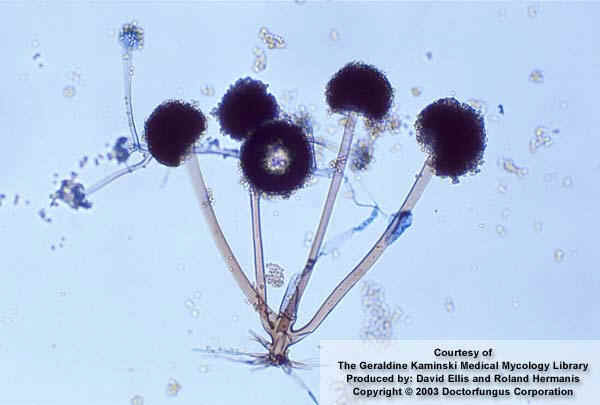
Fungal Growth in Culture (Aspergillus
niger)

 l
l
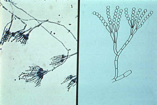
Scopulariopsis,
(S. brevicaulis)
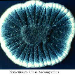
Penicillium
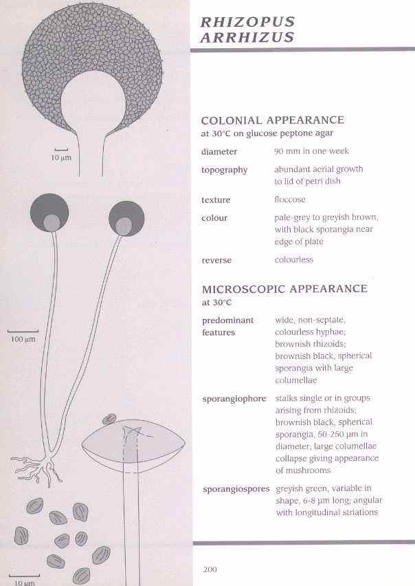
Mucor
Rhizopus
PIEDRA
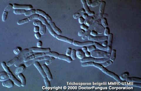
White piedra
(Trichosporon beigelii)
-
white piedra occurs on hair in the beard, axillary regions, groin region,
and less frequently on the scalp.
-
found in temperate regions including the U.S., Europe, Orient, and tropics
(South America).
-
usually associated with poor body hygiene and also in immunosuppressed
patients.
-
pathogen originates from environment, such as soil, stagnant water, spoiling
fruit and food and can be part of the normal skin microflora.
-
If sexual or perfect stage of the fungus is ever discovered, most likely
it will be a basidiomycete due similarity between the ultrastructure of
the hyphal cross walls (dolipore septum is present) of this fungus with
those found in the basidiomycetes.
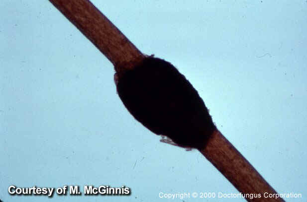
Black piedra
(Piedraia
hortae or P. hortai -
Refer to Figures 34-36 on page 53 in the textbook)
-
caused by an ascomycete that produces stony nodules [stromata] that contain
locules with asci and ascospores)
-
source of infection is soil
Identification
-
make slide using infected hair soaked in 10% KOH solution and examine under
microscope
-
for Piedraia hortae look for locules in the stony stroma and asci
(with 8 ascospores).
-
for Trichosporon beigelii - material detaches easily form hair shaft;
material consists of yeast-like conidia called blastoconidia and a loose
network of hyphae. No ascospores are ever seen.
Both types of piedra are easy to treat!
-
shaving or cutting away infected hair
-
application of topical fungicides ( such as, HgCl2 (1:2000),
benzoic and salicylic acid combinations, 3 percent sulfur ointment or 2
% formalin)
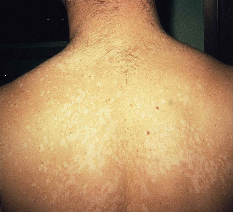
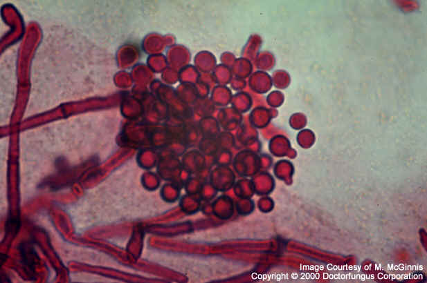
PITYRIASIS VERSICOLOR
-
caused by Malassezia furfur (etiologic or causal agent) and other Malassezia
species
-
Yeast cells occurs in the stratum corneum - compact dead layer of skin
-
cause discovered 150 years ago
-
lipophilic yeast (In culture, olive oil is added to medium)
-
chronic, mild, usually asymptomatic infection of the stratum corneum
-
desquamating (peeling) macules (discolored unraised area of skin)
-
skin cells come off in patches with Scotch tape
-
fairly common and benign disease
-
chest, abdomen, upper limbs, and back
-
on lighter skin, the macules appear dark in color
-
on darker skin, the macules appear light in color
-
exposure with Wood's lamp causes patches to fluoresce yellow to golden
-
look at skin scrapings under the microscope (soak skin scrapings with KOH
solution to clear tissue) - mycelial fragments and budding yeast cells.
-
http://www.doctorfungus.org/mycoses/human/other/Pityriasis_versicolor.htm
-
http://dermnetnz.org/fungal/pityriasis-versicolor.html
Treatment
-
topical application of Whitfield's ointment (contains salicylic acid),
fungicidal agents such as miconazole, and others to a lesser extent.
-
hot soak baths twice a day to remove dead skin.
-
washing clothes with Clorox- sodium hypochlorite to disinfest these.
TINEA NIGRA
-
superficial asymptomatic fungus.
-
infection of the stratum corneum characterized by brown to black non-scalelike
macules often on the palms of hands.
-
etiologic agent - Exophiala werneckii (Sometimes called Cladosporium
werneckii).
-
the word "tinea", refers to ringworm (in this case, not caused by a dermatophyte),
while the word "nigra" refers to black macules associated with this disease.
-
resembles a silver nitrate stain but irregular.
-
sometimes confused with a malignant melanoma (difference being that melanomas
are often raised)
-
primarily a tropical disease (Central and South America, Africa and Asia).
-
examine skin scrapings to find hyphae, which differ from a dermatophyte
(colorless or hyaline hyphae) in being darkly pigmented, often brown to
olivaceous in color.
-
hyphae are highly branched and septate.
Treatment - readily treated, over several weeks
-
Whitfield's ointment, miconazole nitrate in a cream base, as well as other
imidazoles and triazoles applied topically.
-
griseofulvin treatment is not effective
-
sulfur ointments
-
tincture of iodine
Internet Links for
additional information
